Smear for oncocytology. What does squamous epithelium mean in large quantities in a smear? Squamous cells found
Going to the doctor is not a great time for any of us. But, paying attention to our health, we are forced to agree to various unpleasant procedures. Many women think about their visit to the gynecologist with some stiffness and hostility. Ideally, representatives of the fair sex should go to this specialist 2 times a year, but the realities of life are such that this ideal is not available to everyone. Family, work, difficult relationships with someone, a turbulent personal life, failures, stress put off visiting a gynecologist until health problems become acute.
When visiting a gynecologist, you need to be prepared for the fact that you will have to take tests to determine bacteria and the presence of pathogenic microorganisms in the vagina. After receiving the results, many questions arise, for example, whether there should be squamous epithelium in the smear, or what amount of bacteria and other elements is allowed in the flora. This article will discuss the properties, types and quantities of squamous epithelium in the analyses.
Indications for prescribing analysis
Cell testing should be performed regularly on all women over 18 years of age. It is prescribed once a year and does not depend on the health status of the representative of the weaker half of society. If there are any pathological changes in the cervix, the doctor may order tests as long as necessary. Since recently diseases of the female genital organs have become younger, the environmental situation has worsened and people have become more susceptible to stress, experts prefer to prescribe a smear for cell examination at least 2 times a year.
Without this analysis, it is almost impossible to accurately determine the pathological processes occurring in the cervix. is popular because it allows you to quickly and safely identify inflammatory, precancerous and cancerous conditions in a woman. In addition to the fact that you can see squamous epithelial cells in a smear, it also displays the presence of leukocytes, bacteria, and fungus.
Can squamous epithelium be present in a smear?

Sometimes women, when receiving test results, are frightened by the presence of squamous epithelial cells in it. But don’t worry, because their presence is physiologically justified. The fact is that the cervix and vagina are lined with tissue called squamous epithelium. In a smear, the norm of these cells in the field of view is up to 15 pieces. Their absence or a significant deviation from the norm indicates the presence of local pathological processes. You should never draw a conclusion about your health status based only on this indicator in the analysis. A doctor can get a complete picture of a woman’s health (or lack thereof) only by comparing the indicators of squamous epithelium in a smear with other elements.
Squamous epithelium in a smear in small quantities

Low values of any element in analyzes do not always indicate normality. After all, any deviation from it can have a detrimental effect on our health. Flat epithelium in a smear (the norm of which is indicated above) may have values of 1,2,4. A small number of these cells may indicate a lack of estrogen production, and an increased amount of male hormones. If these cells are not visible at all upon careful examination, this indicates that they are atrophied. Their complete absence should alert a specialist, since the death of epithelial cells can lead to the development of a cancerous tumor. In order to confirm this assumption, several more analyzes and studies need to be done, so there is no need to panic with such results.
What to do if the squamous epithelium in the smear is higher than normal?

Experts immediately pay attention to the results of the analysis if squamous epithelial cells in the smear are contained in large numbers. Indicators above 15 are considered a deviation from the norm and may indicate the presence of pathological processes such as inflammation of cervical tissue, the development of a benign tumor (diffuse mastopathy). Also, epithelial cells may indicate primary infertility in young patients.
Nuclear-free “scales” (this is what squamous epithelium looks like) can grow without a focus. This is observed in benign tumors, as well as in the pathological process of hyperkeratosis. Hyperkeratosis is a keratinization disorder in which the responsible organs do not control how much and how squamous epithelium appears. There may also be a lot of it in the smear due to a significant excess of estrogen in the body. In this case, the woman is also at risk of miscarriage. Epithelial cells are carefully studied in order to prevent the development of cancer in the early stages.
Various changes in squamous epithelium in a smear
The results of a routine smear test may lead to additional testing and treatment. This occurs when epithelial cells undergo a quantitative change. must comply with the standard in shape, structure and size.
Flat epithelium in a smear can be combined with cylindrical epithelium. This is not a deviation from the norm if the smear was made in the transition zone (the cervical canal and its vaginal part). Given that the epithelium lines the canal and vagina in several layers, the analysis results may show cells from different layers. Stratified squamous epithelium may also appear in the smear; such results without additional abnormalities in cell structure or size are considered within normal limits.
Don't worry too much if you have modified epithelial cells. This is not reliable evidence that cancer is developing. Squamous epithelial cells that are abnormal in structure and structure may indicate ongoing inflammatory processes, the presence of benign lesions of the cervix, and dysplasia.
How does this type of cell change depending on age?

A woman goes through different stages of development in her life, depending on her age, and internal organs and cells change. The squamous epithelium was no exception (in the smear it is designated as “Ep”). In women of reproductive age, the boundary between the arrangement of cylindrical epithelial cells and flat ones is clearly visible. They have a typical appearance, and the analysis results will be reliable due to their correct location. During life, this clear boundary moves into the cervical canal. In women before and during menopause, squamous epithelial cells are no longer as large as they were before. They become thinner, and a gap appears in the vessels.
Is it necessary to sound the alarm when squamous epithelium appears in layers in a smear?

If your squamous epithelium is located in layers in the smear, then you need to consult a specialist for your own peace of mind. Such results should be analyzed starting with its quantity in the field of view. If the norm is not exceeded, the cells are not changed, there is no reason to panic. After all, flat epithelium lines the vagina and the walls of the cervix in layers. But if the number of cells significantly exceeds the norm, you need to immediately go to the gynecologist to schedule a further examination.
How should you prepare for the analysis?
Since a woman lives on a cycle, she needs to know when it is best to do a vaginal smear. During reproductive age, it is important to calculate the days of menstruation, otherwise the squamous epithelium in the smear may be subject to changes. Women received many erroneous results precisely because of incorrect collection of biomaterial. For those representatives of the fair sex who menstruate, a smear should be taken no earlier than the 5th day of menstruation. In addition, the analysis should be done a maximum of 5 days before the start of menstruation, no later. If sexual intercourse took place, medications were introduced into the vagina, or sanitation was performed, the biomaterial will be ready for collection only after 24 hours.
The material is applied to two glasses with a soft brush or spatula. The results are ready in 5-10 days.
What additional studies are prescribed if the squamous epithelium does not correspond to the norm?
If a single squamous epithelium is detected in the smear, but there are no changes in the cervix, then the analysis is considered normal and does not require any additional examinations or studies. But there are some situations when it is necessary to carefully look at the epithelial cells in an enlarged form. This happens when erosion is suspected and the development of cancer. In this case, colposcopy or cervical biopsy is prescribed. Such studies are carried out by a highly professional specialist, since the patient’s life may depend on the diagnosis as a result of the examination. If moderate to severe damage to the cervix is detected, treatment methods such as cauterization or removal of the affected area are prescribed.
Prevention, regular examination and examination, timely treatment of pathological processes can prolong your life for a long time. Take care of yourself and don't get sick!
Content
A correctly performed smear should contain cells of stratified squamous, cylindrical and glandular epithelium, vaginal flora, mucus, and a moderate amount of neutrophils. The ratio of components and the state of each type of cell allow the doctor to identify early pathology of the female and male genital tract.
What is epithelium
All tissue and organ surfaces are protected by integumentary epithelial cells. Depending on the function of the tissue and the intensity of the mechanical load on it, the lining has a different structure and thickness. The skin exposed to the greatest external influences is covered with stratified squamous keratinizing epithelium. Multi-layering is inherent in the lining of individual sections of the respiratory, digestive, and genitourinary tracts. This is explained by proximity to the external environment and frequency of contact with microbial agents.
Flat
The external genital tract has heterogeneous coverings. The vagina and the outer part of the cervix (exocervix) are lined with stratified squamous epithelium. As it matures, the young (basal) layer (layer) seems to be pushed out from the membrane, changing its cellular shape and size. The cytogram contains flat epithelium of the surface layer - the most mature elements with a small nucleus and abundant cytoplasm. The cylindrical epithelium in a smear in women represents the lining of the internal pharynx, part of the cervical canal.
Glandular
The cervical canal is covered with secretory type epithelial cells (endocervix). They produce mucus, the accumulation of which in the canal creates a kind of plug that protects the uterine cavity from infection. A properly performed smear contains endocervical cells; they make up approximately 10% of the cellular components. But if there is a lot of glandular epithelium, then a consultation with a gynecologist is required to exclude proliferative processes and polyps of the cervical canal.
Cylindrical
The bulk of the smear is squamous epithelial cells. Among them there are small groups of cylindrical lining the narrow transitional area (internal os) of the cervix. The absence of such cellular composition may indicate dysfunction in the production of the hormone estrogen, often of a menopausal nature. Occurs in cystic ovarian lesions. Prismatic cells are the same cylindrical ones, but flattened. They appear in smears of elderly patients and are a sign of atrophic and dystrophic processes.
Normal epithelial cells in a smear

The qualitative and quantitative composition of smears taken for cytology depends on two factors. The first is the condition of the patient’s genital tract. The second is the correctness of taking material for the cytological preparation. The glass should contain cells of the vaginal, cervical, and cervical epithelium in women, and cells of the squamous and urethral epithelium in men. Only in this case will the doctor be able to properly evaluate the diagnostic material.
Among women
Squamous epithelial cells predominate in a cytology smear, but one field of view should not contain more than twenty units. The presence of several less mature intermediate cells from the middle (granular) layer is considered the norm. About 10% of the cellular composition is cylindrical and glandular components. The flora is represented by rod-shaped and coccal bacteria; the predominance of one or another variety depends on the phase of the menstrual cycle. The presence of single neutrophils is acceptable.
In men
Normal laboratory tests in men include stratified squamous and urethral cell types. The lining of the urethra is multi-row, there is no division into layers (as in the cervix). For this reason, the urethral component is represented by identical cellular elements - prismatic transitional type. Urine inclusions (a few salt crystals) may be present. Single cocci are acceptable, no more than five elements of the inflammatory series (neutrophils, leukocytes).
What does a large amount of epithelium in a smear mean?

A normal cytogram in a smear for flora contains 12-20 epithelial cells per field of view. Excessive content of the squamous cell component indicates irritation and accelerated rejection of the integumentary layer. The cause may be inflammatory processes of various etiologies, then the doctor will see a significant number of leukocytes in the drug (normally no more than five). Often a pathogen is detected: Trichomonas, viral inclusions.
In the absence of inflammatory elements, one should think about leukoplakia and other types of dyskeratosis. Allergic reactions to local medications (contraceptive drugs, medicinal ointments, suppositories) show a similar picture. Moderate irritation is often caused by hygiene products. The epithelium of the cervix during pregnancy may be somewhat more abundant, the cellular cytoplasm has signs of decidual metamorphosis, this is a normal variant.
The cytological picture in men varies; with age, the squamous cell component can be increased, but its number should not exceed 15 units in one visual field. The abundance of epithelial masses, mucus impurities, and leukocytes indicate an inflammatory process. You should not attempt treatment on your own, as this may lead to the symptoms subside without eliminating the cause of the disease.
Attention! The information presented in the article is for informational purposes only. The materials in the article do not encourage self-treatment. Only a qualified doctor can make a diagnosis and give treatment recommendations based on the individual characteristics of a particular patient.
Found an error in the text? Select it, press Ctrl + Enter and we will fix everything!Often, when visiting a gynecologist, after an examination in a gynecological chair and an ultrasound, a woman receives a referral for a smear for cytology. Let us consider this type of research in detail, name the indications for its implementation, and the features of collecting material.
What is “cytology” in gynecology?
In most cases, when cervical cytology is prescribed, the girl does not know what it is. To begin with, it must be said that the cervical canal is an anatomical formation that in appearance resembles a tube. It directly connects the vagina to the uterine cavity. It contains stratified and columnar epithelium.
Taking material from this area of the reproductive system helps to identify atypical cellular formations in a timely manner. This makes it possible to diagnose a precancerous condition and prescribe appropriate treatment. Evaluation of cellular structures occurs under a microscope with high magnification. The nuclear contents of the cells are subjected to careful analysis.
What does a cytology smear show?
A common option for this type of test is the Pap test. It was first used at the beginning of the 20th century by the Greek scientist Papanikolaou. He was directly involved in the diagnosis of malignant processes in the early stages. This cytology smear also makes it possible to identify background processes of non-tumor origin that have a risk of developing into cancer.
Taking into account all the above possibilities of the PAP test, doctors prescribe it for the following changes in intraepithelial structures:
- abnormalities in the structure of cells of high and low severity;
- the presence of pathogenic microflora in high concentrations as a result of previous studies;
- disturbances in the menstrual cycle of unknown nature;
- diseases of the reproductive system of a viral nature (HPV, herpes);
- abnormal discharge from the vaginal cavity after menstruation.
Atrophic type of smear for cytology
The PAP test in gynecology is one of the main studies that helps identify the cellular ratio in the cervical canal. With the atrophic type of smear, in the field of view of the microscope, the laboratory assistant records a large content of squamous epithelial cells. This indicates changes in the structure of epithelial tissue, which is often recorded in dysplasia. Further diagnostics, by carrying out, establishes the degree and severity of the disorder. A smear for pathological cytology is the initial stage of a comprehensive examination of the reproductive system.

Inflammatory type of smear for cytology
Cytology of the cervix helps to identify inflammatory processes at an early stage and prescribe the correct therapy. With an inflammatory type of smear, changes are recorded at the cellular level - the ratio of glandular cells to squamous epithelial cells may increase. At the same time, the woman notes the presence of symptoms of inflammation, which are manifested in changes in the cycle - acyclic discharge, an increase in the volume of menstrual blood, strong.
When to take a smear for cytology?
Almost like any study of the reproductive system, cytology analysis is carried out on a certain day of the menstrual cycle. The procedure for collecting material is carried out 10-12 days from the moment it begins. It must be taken into account that the result may be inaccurate if there is an infectious process in the body. If necessary, the analysis is carried out even during illness, but after 2 months from the moment of recovery, a control study is prescribed. If a woman uses vaginal suppositories, performs baths, douches, analysis is possible 7 days after completion.
As for the indications for the study, doctors can prescribe the test for:
- frequent childbirth (3 times in 4 years, for example);
- early first childbirth (birth of the first child before 18 years of age);
- placement of an intrauterine device;
- lack of examinations of the reproductive system over the past 3 years;
- the presence of visible changes when examined in a gynecological chair using mirrors;
- burdened medical history (tumor-like processes were recorded in the family on the female side).
Preparing for a smear for cytology
Liquid cytology involves assessing the material after immersing it in a special medium. Previously, a smear for cytology was carried out with the application and fixation of material on glass. To obtain an accurate result that reflects the condition of the uterine structures, a woman must adhere to the following rules before the procedure:
- exclusion of douching 5-7 days before sampling;
- exclusion of sexual intercourse for 3 days;
- do not use tampons, gels, or vaginal creams;
- do not urinate 2 hours before the procedure.

How is a smear taken for cytology?
The Pap smear is performed in a clinic setting. The woman sits in a gynecological chair. During the procedure itself, the gynecologist takes cells from the cervical canal area, from the vaginal mucosa. In the first case, a special probe is used - endobrush. It is administered after a slight expansion of the uterine canal by using a dilator. An Eyre spatula is used to collect material from the vaginal walls.
When liquid cytology of the cervix is performed, during the manipulation itself the woman may feel minor discomfort and mild pain. Its appearance is due to the expansion of the cervical canal, which is supplied with a large number of nerve endings. The duration of the procedure depends on the experience of the gynecologist, and averages 5-10 minutes. The resulting material is placed in a test tube with a reagent and sent to the laboratory.
Cytology smear - interpretation, normal
After a cytology smear has been performed, the results are deciphered exclusively by a doctor. Only a specialist can perform a comprehensive analysis of the situation by assessing the results of cytology. The state of cellular structures is a reflection of the state of the reproductive system. Based on the results obtained, doctors can suggest an oncological process and prescribe additional tests (colposcopy, curettage).
Atypical cells in a smear for cytology
When a woman learns that the results of the study revealed she has poor cytology, this news becomes a cause for concern. It is worth noting that such a conclusion does not mean cancer. According to the established terminology of medical reports, this definition indicates the presence of pathological changes in the epithelial layer of the vagina and cervical canal.
According to the generally accepted methodology for assessing results, changes are designated as follows:
- 0 – the collected material is of low quality, unsatisfactory (selected in small quantities, the patient was not properly prepared);
- 1st class – indicators are normal;
- Class 2 – atypical structures are present;
- Class 3 - there is dysplasia of varying severity;
- Class 4 – precancerous condition, first stage;
- 5th grade – cancer.

Squamous epithelial cells in a smear for cytology
When liquid cytology of the cervix diagnoses squamous epithelial cells in the canal, doctors talk about a disease such as hyperkeratosis. A large number of squamous epithelial scales are recorded in the field of view of the microscope. This type of cellular structures is present in analyzes with. This disease is a benign tumor. When conducting an additional study, colposcopy, a white area is recorded on the cervix.
It is worth noting that when the smear results contain only single scales, colposcopy is mandatory. If during its implementation no changes are revealed on the surface of the reproductive organ, then the analysis is considered normal. A cytology smear performed in this case has no diagnostic value. The patient is monitored - once every 3 months the woman visits the consultation department and is examined.
Glandular epithelium in a smear for cytology
Analysis of the smear for cytology, its interpretation, further helps to establish inflammatory processes in the uterus. One of these is cervical dysplasia. With a mild degree of the disorder, the disease can be easily corrected by prescribing anti-inflammatory drugs. Moderate and severe dysplasia can be regarded as a precancerous condition. In this case, a biopsy becomes a mandatory additional study - taking a section of the affected cervical tissue for histological examination.
Polymorphic rod flora in a smear for cytology
Cytology transcripts often contain the entry “polymorphic rod flora.” In order to understand what this means, it is necessary to say that the rods present in the vagina may have:
- morphotype of lactobacilli (similar in structure and appearance to Doderlein sticks);
- small sticks.
The first type of rods makes up the normal microflora of the vagina. At the same time, the presence of single leukocytes is allowed. The presence of a large number of small rods indicates a change in the composition of the microflora, which can be recorded with vaginal dysbiosis, gardnerellosis. Such conditions require urgent medical intervention and therapy.
Smears for cytological examination are obtained from the surface of the mucous membrane, therefore their cellular composition is represented by desquamated cells located on the surface of the epithelial layer. The better the epithelium’s ability to mature, the more mature cells appear in the smear; with atrophic changes, less mature cells are located on the surface of the epithelial layer.
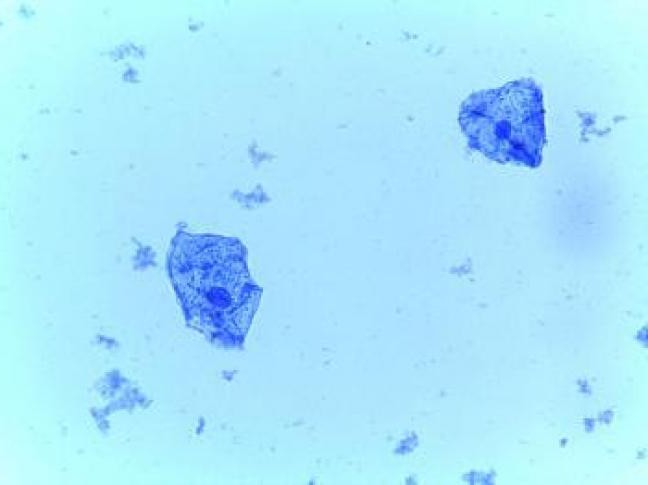
Superficial cells are large flat polygonal, about 50 µm in diameter. The nuclei are oval or round, structureless, pyknotic with a maximum diameter of 5–6 μm. Mature cells are located predominantly scattered, the cytoplasm when stained with Papanicolaou is pinkish-yellow, eosinophilic, and tender. Transparent, in some cells lipid granules and glycogen granules are detected (Fig. 12, a). Less mature cells can be arranged in layers and piled on top of each other. The cytoplasm is cyanophilic, delicate, transparent, with folds, its contours are clear and uneven.
Intermediate cells are relatively large, usually polygonal. The nuclei are vesicle-shaped, with a clear chromatin structure, more than 6 µm in diameter (Fig. 12, b). The cytoplasm can be eosinophilic, cyanophilic, and folding is characteristic (Fig. 13, a, b). Mature intermediate cells (prepyknotic) differ from superficial ones in the size and structure of the nucleus.
|
|
|
|
|
Rice. 13. Squamous epithelial cells: a) - mature intermediate cells with oval and round vesicular nuclei. |
||


The cytoplasm is abundant, polygonal, green in color. The nuclei are round, the uniformly granular chromatin structure is clearly visible. Papanicolaou staining. x 500; b) - a superficial cell with orange cytoplasm and a pyknotic nucleus (1) and an intermediate cell with a vesicular nucleus, light cyanophilic cytoplasm (2), a few leukocytes. Ectocervix smear.
Papanicolaou staining. x 500
Less mature intermediate cells (navicular, scaphoid) are oval in shape, smaller in size, and their cytoplasm is more dense. Lactobacilli are capable of causing lysis of intermediate cells: this peptic effect rarely extends to superficial cells.
Columnar epithelial cells are normally located in small groups, in the form of stripes, honeycomb-like structures. The cells are elongated, the nuclei are located eccentrically. There may be “goblet” cells in which the cytoplasm is stretched with mucus; sometimes secretion granules are found in the cells (Fig. 15 – 17).
|
|
|
|
|
Rice. 15. Columnar epithelial cells in the form of a honeycomb-like structure. The nuclei are round or oval, the contours are smooth, the chromatin is evenly distributed, and small monomorphic nucleoli are found. The cytoplasm is vacuolated. |
Cervical smear. |
|
|
|
|
|
|
Staining according to Romanovsky. x 1000 |
||


Rice. 16. A small group of columnar epithelial cells. The nuclei are oval, the contours are smooth, the chromatin is granular and evenly distributed.
In the cytoplasm there are small secretion granules.
Cervical smear. Staining according to Romanovsky. x 1000
Rice. 17. Smear from the endocervix: a) - a small structure of columnar epithelial cells in the form of a strip (1), squamous epithelial cells of the surface (2) and intermediate (3) layers, erythrocytes.
Papanicolaou staining. x 400 b) - a small group of columnar epithelial cells. Staining according to Romanovsky.
x 400
Almost every woman knows that squamous epithelium in a smear is a very important indicator with which a doctor can determine the condition of the mucous layer of the genital organs.
Cytology analysis provides reliable information about the hormonal background, as well as the presence or absence of a pathological process in the genitourinary system.
Very often, patients ask the question of whether squamous epithelial cells should be present in the smear and how many of them there should be, after receiving the result of the cytology test.
The epithelial layer consists of superficial, basal and intermediate cells. Every six to seven days (as it matures), the young layer changes its cellular shape and size.
Obsolete particles are peeled off and replaced with new ones, so squamous epithelium should be present in smears of healthy women.
Due to the formation of new cells, the thickness of the mucous membrane increases. It should be noted that squamous epithelium consists of elements with small nuclei and abundant cytoplasm.
The gynecologist attaches great importance to the results of the analysis. When deciphering, the doctor analyzes the condition of the genital organs and draws conclusions about the presence or absence of hormonal imbalance.
An insufficient number or a significant increase in the cells that form the mucous layer indicates diseases of the body that can lead to the formation of tumors.
The presence of epithelial particles in the smear may indicate vaginitis, urethritis, a lack or excess of certain hormones. Only an analysis will tell you whether such cells are dangerous or not.

The epithelial cover contains cells that make up the intermediate layer. The thickness of the squamous epithelium is one hundred fifty to two hundred microns.
In addition, the smear may contain cylindrical epithelial cells that line the internal os and partially the cervical canal.
The covering provides the necessary amount of mucus to lubricate the cervix. The indicator of cylindrical epithelial cells should also correspond to the norm.
Why is a smear necessary?
Most women pay their visit to the gynecologist with some constraint, modesty or even hostility.
Avoiding troubles for women in terms of health is quite simple.
It is enough to regularly take a cytology test, the results of which can provide answers to many questions. A smear can tell about the presence of not only bacteria, fungi or leukocytes.
The result of the smear analysis will help determine the number of epithelial cells and promptly identify inflammatory (sometimes even cancerous or precancerous) processes in a woman.
The gynecologist definitely recommends taking a cytology test (PAP test) at least once a year. When planning a pregnancy, this analysis is inevitable.
It is very important to get the result of a smear for pregnant women who did not have time to take it in the process of planning a future child.
During pregnancy, the flat epithelium that lines the vagina is capable of retaining harmful microorganisms.
By detecting an increased number of epithelial cells in time, the doctor will help avoid the development of a severe inflammatory process in the expectant mother. Lack of estrogen in a pregnant woman can be easily diagnosed by a low level of squamous epithelium.
Experts prescribe this test to women who want to have an intrauterine device installed, if they suspect the presence of genital herpes, in cases of infertility or menstrual irregularities.
Patients who are overweight (obese) may have gynecological disorders, and the doctor also often recommends a smear test.
The best time to conduct the study is the fourth or fifth day of the cycle. Before taking the test, a woman should abstain from sexual intercourse for at least two or three days, avoid the use of ointments, lubricants, and douching.
You need to stop going to the toilet two to three hours before visiting the gynecologist. Before this, you first need to do a hygienic wash.
A smear is taken from women during a gynecological examination using a special small disposable brush. The material is carefully taken from the surface of the cervix.
The procedure is painless, but at the time of taking the scraping, slightly unpleasant sensations may occur.
After the doctor takes the material for analysis, some women may experience slight spotting. This is a normal phenomenon, the patient should not be afraid of this.
Normal squamous epithelium in a smear
The microbial composition of the smear is determined by specialists in laboratory conditions. With the help of special reagents, the epithelium in the scraping is painted in different colors, with the help of which you can designate and evaluate the composition of the material taken for analysis.
The results of the smear are usually known within one day. When all particles of squamous epithelium have the correct shape and size, the analysis is considered normal. There must be no atypical cells.
If particles with pathology are detected, the doctor must prescribe additional studies of the reasons for their appearance.

As a result of the analysis, a woman may see the abbreviation “Ep”, which means epithelium. In a smear, the normal number of squamous epithelial particles is about fifteen pieces.
A deviation in any direction indicates that some local pathologies are developing in the woman’s body that threaten her health. If the result of the presence of squamous epithelium is overestimated, then the patient may exhibit inflammatory processes.
A woman of childbearing age with an increased deviation from the norm in a smear may suffer from infertility. Often, an increase in the amount of squamous epithelium indicates the presence of vaginitis.
Patients often visit a gynecologist with complaints of itching in the vaginal area, the presence of copious unpleasant discharge, sometimes with a characteristic odor.
In such cases, the doctor prescribes appropriate antibacterial drugs. To avoid re-infection, it is recommended that the sexual partner of the sick patient also undergo examination.
A situation in which the amount of squamous epithelium in a scraping is significantly higher than normal may be a sign that a woman has gardnerella, trichomonas, gonococcus and other pathogens.
One of the reasons for the presence of a large number of epithelial cells in a smear may be estrogens.
Excessive saturation of the body with hormones of this group can lead to termination of pregnancy.
An underestimated amount of squamous epithelium in a smear is also an alarming symptom, since it indicates thinning of the vaginal mucous membranes.
Experts attribute the decrease in the indicator to estrogen deficiency in the patient. Women, as a rule, note an almost complete absence of discharge.
During sex, an insufficient amount of lubrication is produced, which makes sexual intercourse much more difficult. A small number of epithelial cells usually alerts doctors.
There is an assumption that a woman with this result has a tendency to develop cancerous tumors.
Timely diagnosis of deviations from the norm will help to avoid the further development of a large number of diseases in women. Do not neglect visiting a gynecologist.
At the slightest discomfort, a woman should try to visit a doctor as soon as possible and take a cytology test.
It should be noted that after proper treatment, the squamous epithelium in the scraping often takes on normal values.